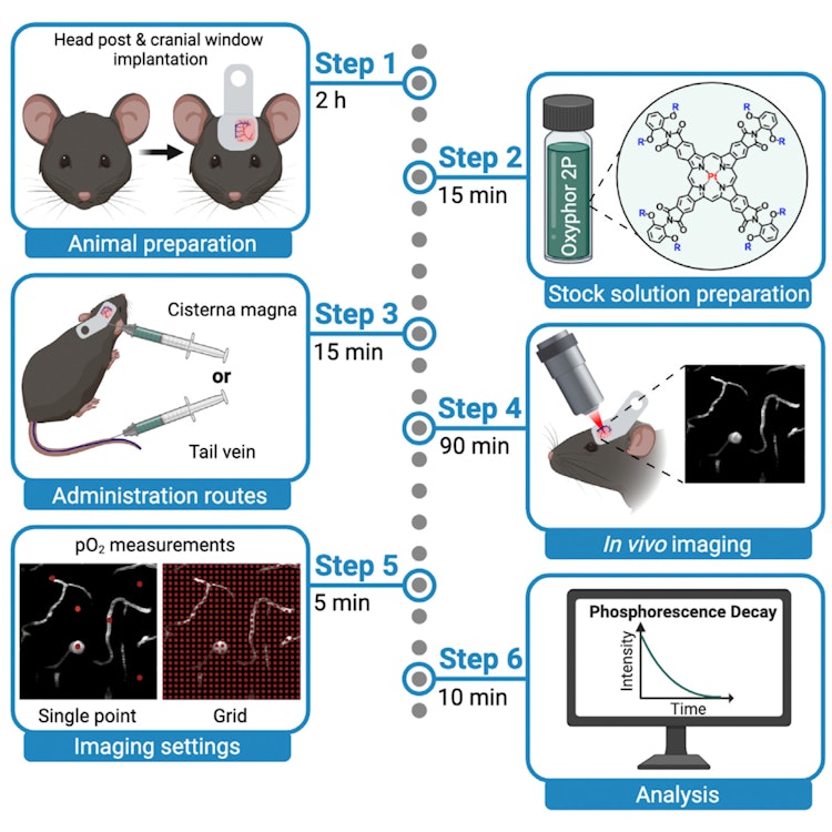
Measurement of cerebral oxygen pressure in living mice by two-photon phosphorescence lifetime microscopy
The ability to quantify partial pressure of oxygen (pO2) is of primary importance for studies of metabolic processes in health and disease. Here, we present a protocol for imaging of oxygen distributions in tissue and vasculature of the cerebral cortex of anesthetized and awake mice. We describe in vivo two-photon phosphorescence lifetime microscopy (2PLM) of oxygen using the probe Oxyphor 2P. This minimally invasive protocol outperforms existing approaches in terms of accuracy, resolution, and imaging depth. For complete details on the use and execution of this protocol, please refer to Esipova et al. (2019).
Download
erlebach_2022.pdfResearchers
Eva Erlebach

Dr. Luca Ravotto

PD Dr. Dr. Matthias T Wyss

Jacqueline Condrau
Thomas Troxler
Sergei A Vinogradov

Prof. Dr. Bruno Weber


